
While conventional lasers rely strictly on selective photothermolysis via chromophores (melanin, hemoglobin, water), their application in darker skin phototypes and melasma carries severe risks of post-inflammatory hyperpigmentation (PIH) and irreversible depigmentation (mottled hypopigmentation) from excessive laser toning. Furthermore, conditions such as atrophic acne scars, rosacea, and recalcitrant melasma are not merely epidermal; they stem from basement membrane degradation, dermal photoaging, and abnormal microvascular proliferation.
In this webinar summary, Dr. Utako Kimura elucidates the thermodynamic principles of chromophore-independent Joule heating, histological biopsy evidence proving a 3-month ECM and elastic fiber remodeling cycle, and pathology-guided Microneedle RF protocols targeting the basement membrane and microvascular environment.
Radiofrequency Thermodynamics Beyond Lasers & Temperature-Dependent Tissue Alterations [01:02~]
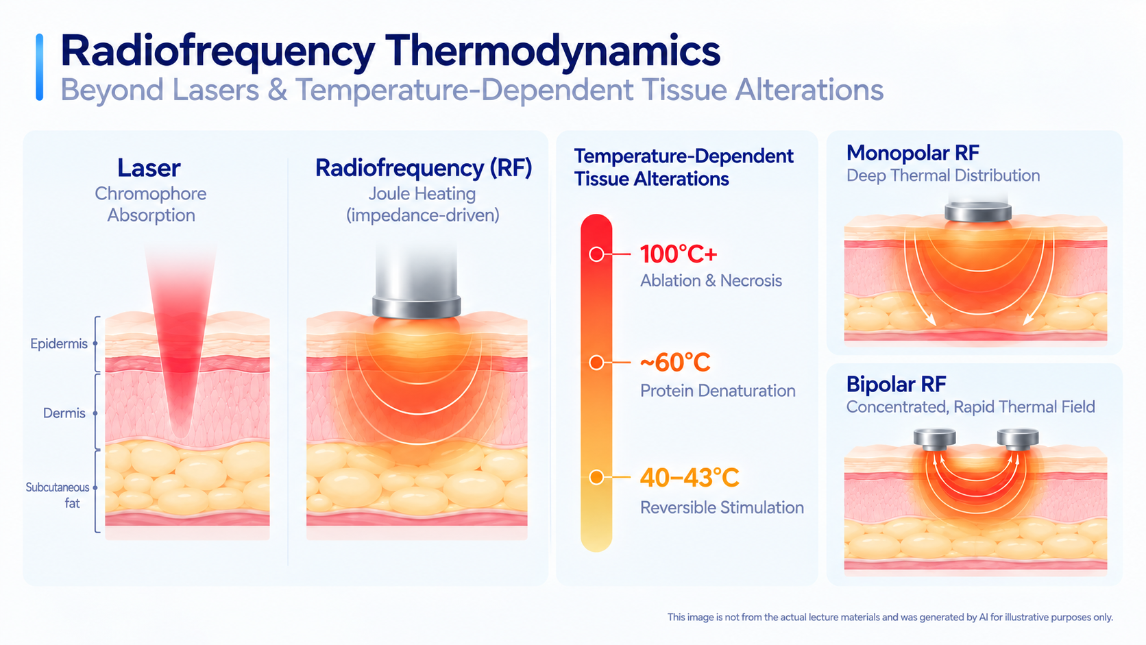
Unlike light-based lasers that depend on chromophore absorption, radiofrequency generates pure thermal energy through Joule heating—the resistance (impedance) of tissue to high-frequency electromagnetic currents. This mechanism preferentially heats fibrous connective tissue over subcutaneous fat or water, avoiding the epidermal melanin interference that complicates laser use in darker phototypes.
Tissue outcomes are strictly governed by temperature thresholds:
- 40–43°C (Reversible Stimulation): Induces heat shock proteins and cytokines, initiating gradual collagen synthesis.
- 60°C (Critical Protein Denaturation): Reaches the exact boundary of reparable irreversible injury, triggering immediate tissue coagulation and contraction for profound dermal tightening without surface destruction.
- 100°C+ (Ablation & Necrosis): Causes vaporization, requiring strict parameter boundaries.
Monopolar modes diffuse deeply toward a return grounding pad, whereas Bipolar modes generate concentrated, rapid thermal fields between closely paired electrodes in the upper dermis.
Biopsy-Verified Dermal ECM & Elastic Fiber Remodeling Timeline [12:28~]
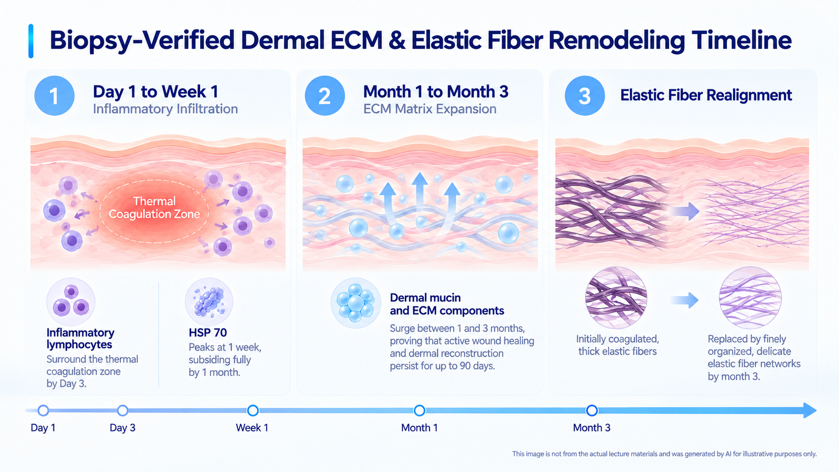
Histological evaluations of ex-vivo human abdominal skin biopsies across HE, Colloidal Iron, and EVG stains confirm that needle insertion alone cannot denature collagen; focused RF thermal zones are essential to drive true three-phase tissue remodeling:
- Day 1 to Week 1 (Inflammatory Infiltration): Inflammatory lymphocytes surround the thermal coagulation zone by Day 3, while Heat Shock Protein 70 (HSP 70) peaks at 1 week, subsiding fully by 1 month.
- Month 1 to Month 3 (ECM Matrix Expansion): Colloidal iron staining reveals that dermal mucin and extracellular matrix components surge between 1 and 3 months, proving that active wound healing and dermal reconstruction persist for up to 90 days.
- Elastic Fiber Realignment: EVG staining demonstrates that initially coagulated, thick elastic fibers are progressively replaced by finely organized, delicate elastic fiber networks by month 3, providing the structural basis for pore refinement, wrinkle reduction, and scar correction.
Melasma Paradigm Shift: Overcoming Toning Limitations via Basement Membrane Repair [20:36~]
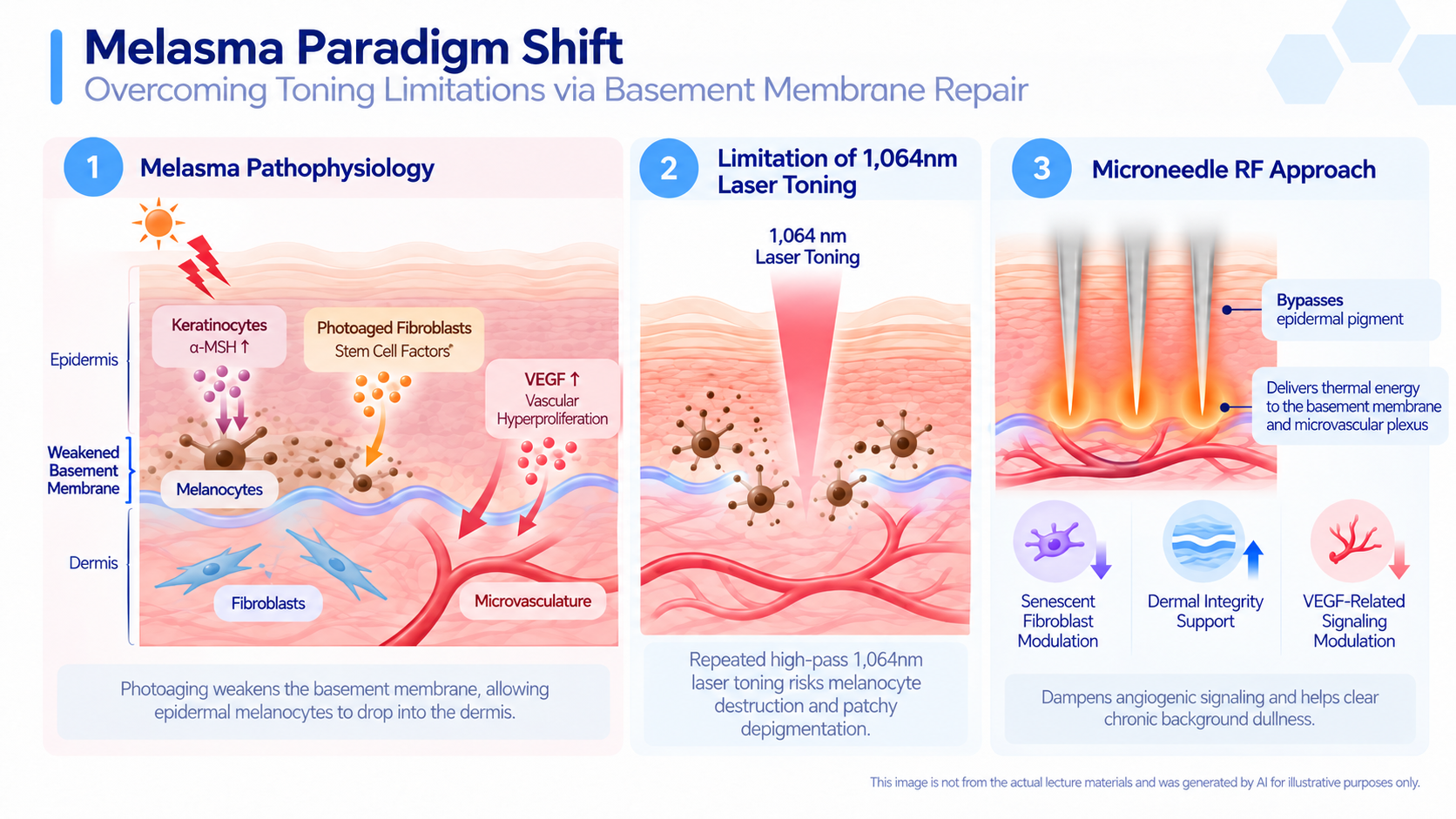
Melasma is an inflammatory storm involving keratinocyte-derived α-MSH, photoaged fibroblasts releasing stem cell factors, and elevated VEGF promoting vascular hyperproliferation. Crucially, photoaging weakens the basement membrane, allowing epidermal melanocytes to drop into the dermis. In this state, repeated high-pass 1,064nm laser toning risks melanocyte destruction and patchy depigmentation.
Microneedle RF provides a safer, restorative alternative by bypassing epidermal pigment to deliver controlled thermal energy directly to the basement membrane and microvascular plexus. This reduces senescent fibroblasts, restores dermal integrity, and calms vascular hyperactivity. For melasma patients unresponsive to toning, as well as those suffering from rosacea erythema, Microneedle RF dampens angiogenic signaling (VEGF) and clears chronic background dullness
Pathology-Specific Tip Selection (S, CP, Diamond) & Real-World Clinical Efficacy [26:26~]
Clinical success depends on matching specific needle architectures and energy modes to anatomical target depths:
- S Tip (Alternating Insulation Pattern): Calibrated for superficial dermal and basement membrane remodeling, effectively treating enlarged pores, fine periorbital lines, rosacea erythema, and recalcitrant melasma.
- CP Tip (Negative-Pressure Drug Delivery): Unites needle stimulation, RF coagulation, and transdermal suction. Delivering the PLLA agent McCoom across 5 sessions achieved substantial dermal filling of atrophic acne scars, marked pore reduction, and resolved post-inflammatory erythema (PIE).
- Diamond Tip (Non-Invasive RF): Employs non-penetrating surface electrodes for zero-downtime, needle-free tightening and textural radiance.
- One Needle (Single-Needle Coagulation): Delivers pinpoint destruction for active acne lesions and syringomas.
By anchoring treatment in precise anatomical diagnosis and customized tip depth, practitioners can safely reconstruct the dermis while avoiding post-inflammatory complications.